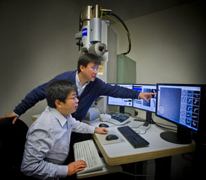
An international team of staff and users working at the Molecular Foundry has captured the first high-resolution 3D images from individual double-helix DNA segments attached at either end to gold nanoparticles. The images detail the flexible structure of the DNA segments, which appear as nanoscale jump ropes.
This unique imaging capability developed at the Foundry is called individual-particle electron tomography (IPET) and could aid in the use of DNA segments as building blocks for molecular devices that function as nanoscale drug-delivery systems, markers for biological research, and components for computer memory and electronic devices. It could also lead to images of important disease-relevant proteins that have proven elusive for other imaging techniques, and of the assembly process that forms DNA from separate, individual strands.
The shapes of the coiled DNA strands, which were sandwiched between polygon-shaped gold nanoparticles, were reconstructed in 3D using a cutting-edge electron microscope technique coupled with a protein-staining process and sophisticated software that provided structural details to the scale of about 2 nanometers, or two billionths of a meter.Read the full press release.

