Scientific Achievement
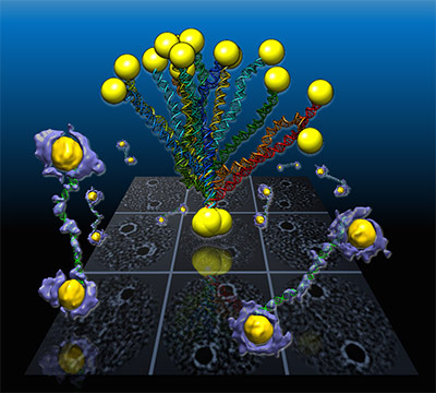
An international team of staff and users working at the Molecular Foundry has captured the first high-resolution 3D images from individual double-helix DNA segments attached at either end to gold nanoparticles
Significance and Impact
This unique imaging capability should lead to better understanding of disease-relevant proteins and the assembly process that forms DNA. It could also aid in the use of DNA segments as biomarkers, drug-delivery systems, computer memory and electronic devices.
Research Details
- Developed at the Foundry, the imaging technique is called individual-particle electron tomography (IPET). IPET takes 2D images from different angles of the same object, which is flash-frozen to preserve the structure during cryo-EM imaging, to assemble a 3D image.
- The shapes of the coiled DNA strands, which were sandwiched between polygon-shaped gold nanoparticles, were imaged using IPET, with help from a protein-staining process and molecular simulation tools that provided structural details to the scale of about 2 nm.
- The molecular modeling simulated natural shape variations, called “conformations,” in the samples, and compared these shapes with observations.

