By Brooke Kuei

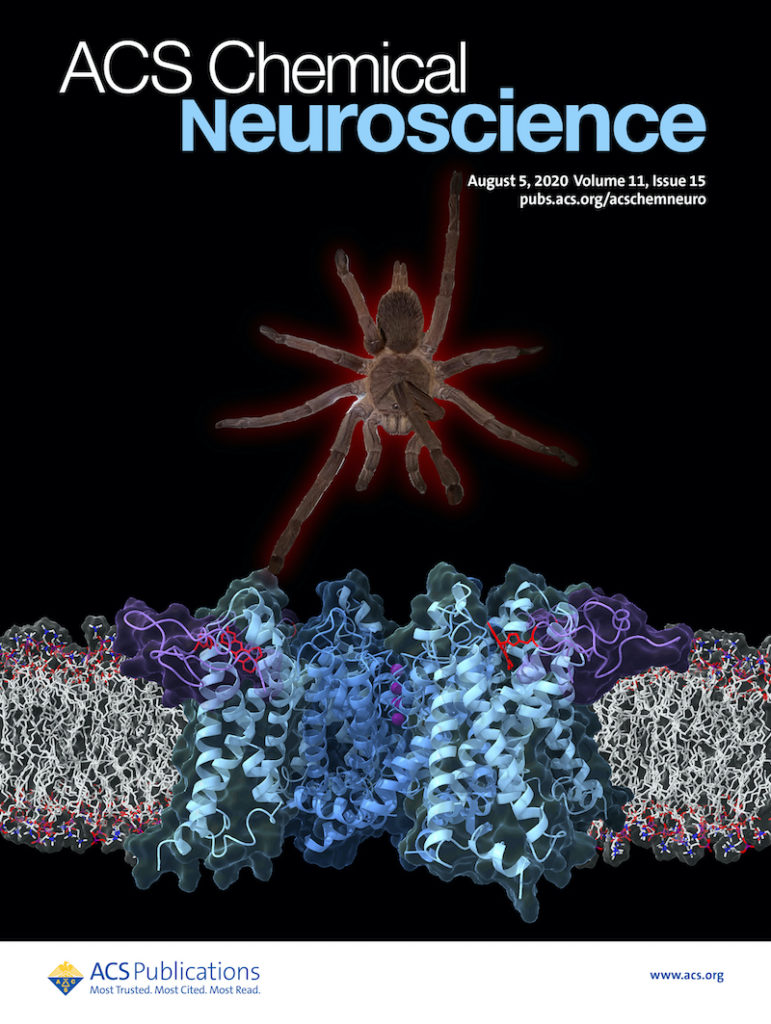
When Jon Sack, associate professor at UC Davis and Molecular Foundry user, brought his lab’s pet tarantula Lucy for a visit to the Molecular Foundry, the fuzzy arachnid was met with some trepidation. Now, Lucy is the star of a new journal cover for ACS Chemical Neuroscience featuring a collaboration with the Foundry that enables imaging of different ion channel structures, or conformations, in a live cell.
Ion channels, proteins in cell membranes that allow ions to flow in and out of a cell, play an important role in how our brains and hearts function. The complex ways in which they change shape in a cell membrane, however, make them difficult to image with conventional methods. In this recent work, a tarantula toxin combined with an environment-sensitive chemical is used to image the different conformations of an ion channel in a live cell.
The researchers targeted a voltage-gated potassium channel, which moves potassium ions in and out of the cell in response to changes in voltage. In other words, the channel will open and close based off how positive and negative ions are distributed on either side of the cell membrane.
“Transistors allow a computer chip to process information, and these voltage gated channels act like transistors to allow us to think,” explained Sack. But because the way they change conformations is only relevant in the context of a live cell membrane, it is difficult to capture the movement of ion channels using methods such as X-ray crystallography or cryo-EM.
It turns out that tarantulas and other venomous predators have evolved toxins so that they specifically target voltage-gated potassium channels, making them unique tools that researchers can use to anchor probes onto these channels.
“The tarantula toxin preferentially binds to the closed state of the particular potassium channel we’re studying,” said Bruce Cohen, staff scientist in the Biological Nanostructures facility and a co-author of the project. “From our point of view, we have a way to now use the tarantula’s clever chemistry to understand how these channels are opening and closing.”
The researchers have made a colorful version of a synthetic tarantula toxin by attaching it to an environment-sensitive fluorophore, a chemical that emits, or gives off, different colors of light depending on its surroundings. When the fluorophore is in water, it will emit red light, whereas if it is in a greasier environment like a lipid membrane, it will emit closer to yellow or orange. This results in a probe that specifically targets voltage-gated potassium channels in live cells and changes color when the channel changes its conformation.
This live-cell imaging method illuminates a series of conformation changes, and these minor but important changes can now be directly imaged using the tarantula toxin linked to the environment-sensitive fluorophore.
Moving forward, the scientists are developing more probes to understand different kinds of ion channels and delve deeper into characterizing their structures. They will also use their newly developed tools to understand the role of these channels in neurons, such as in functions like developing memories and signaling that can lead to pain.
More recently, the fluorophore in this work is also being adapted to detect COVID-19 on surfaces. By attaching the fluorophore to peptides that bind to the SARS virus, it is possible to develop a sensor for detecting SARS viruses on surfaces.
“We are using what we know about fluorescent molecules to design sensors for SARS,” said Cohen. “It’s a very different system, but we have been working with tiny parts of the virus and showing that we can see color changes.”

