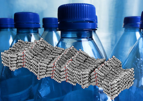
From water bottles and food containers to toys and tubing, many modern materials are made of plastics. And while we produce about 110 million tons per year of synthetic polymers like polyethylene and polypropylene worldwide for these plastic products, there are still mysteries about polymers at the atomic scale.
Because of the difficulty in capturing images of these materials at tiny scales, images of individual atoms in polymers have only been realized in computer simulations and illustrations, for example.
Now, a research team led by Foundry users has adapted a powerful electron-based imaging technique to obtain an image of atomic-scale structure in a synthetic polymer. The research could ultimately inform polymer fabrication methods and lead to new designs for materials and devices that incorporate polymers.
In their study, published in the American Chemical Society’s Macromolecules journal, the researchers detail the development of a cryogenic electron microscopy imaging technique, aided by computerized simulations and sorting techniques, that identified 35 arrangements of crystal structures in a peptoid polymer sample. Peptoids are synthetically produced molecules that mimic biological molecules, including chains of amino acids known as peptides.
The sample was robotically synthesized at Berkeley Lab’s Molecular Foundry, a DOE Office of Science User Facility for nanoscience research. Researchers formed sheets of crystallized polymers measuring about 5 nanometers (billionths of a meter) in thickness when dispersed in water.
The research team created tiny flakes of peptoid nanosheets, froze them to preserve their structure, and then imaged them using an electron beam. An inherent challenge in imaging materials with a soft structure, such as polymers, is that the beam used to capture images also damages the samples.
The direct cryogenic electron microscopy images, obtained using very few electrons to minimize beam damage, are too blurry to reveal individual atoms. Researchers achieved resolution of about 2 angstroms, which is two-tenths of nanometer (billionth of a meter), or about double the diameter of a hydrogen atom.
They achieved this by taking over 500,000 blurry images, sorting different motifs into different “bins,” and averaging the images in each bin. The sorting methods they used were based on algorithms developed by the structural biology community to image the atomic structure of proteins.

