Date: Tuesday, April 27, 2021
Time: 11:00 am
Talk Title: High-Speed Atomic Force Microscopy: A forceful Tool for Molecular Biophysics
Watch the recording

Abstract:
High-speed atomic force microscopy (HS-AFM) is a powerful technique that provides dynamic movies of biomolecules at work. HS-AFM has the notable advantage that it permits to modulate the applied force during imaging, thus representing an excellent tool to study mechanosensitive molecules. Piezo proteins are mechanosensitive ion channels that mediate force-detection in eukaryotic cells through translating a mechanical stimulus into an electrical signal. Recent cryo-EM studies have revealed the structure of most parts of the channel, and gating mechanisms have been suggested. However, it is intrinsically difficult to acquire a structural view of the channel exposed to force. Using HS-AFM imaging during force-sweep cycles, we show that Piezo1 undergoes significant conformational changes under force: the channel reversibly flattens into the membrane plane. In the framework of a model where channel flattening relates to in-plane area expansion, we also derive from the HS-AFM experiments the sensitivity of Piezo1 [1].
To break current temporal limitations to characterize molecular dynamics using HS-AFM, we developed HS-AFM height spectroscopy (HS-AFM-HS), a technique whereby we oscillate the HS-AFM tip at a fixed position and detect the motions of the molecules under the tip. This gives sub-nanometer spatial resolution combined with microseconds temporal resolution of molecular fluctuations. HS-AFM-HS can be used in conjunction with HS-AFM imaging modes, thus giving access to a wide dynamic range [2].
To break current resolution limitations, we developed Localization AFM (LAFM). By applying localization image reconstruction algorithms to peak positions in high-speed AFM and conventional AFM data, we increase the resolution beyond the limits set by the tip radius and resolve single amino acid residues on soft protein surfaces in native and dynamic conditions. The LAFM method allows the calculation of high-resolution maps from either images of many molecules or many images of a single molecule acquired over time, opening new avenues for single molecule structural analysis [3].
[1] Lin et al. Nature, 2019, 573(7773):230–234, Force-induced conformational changes in Piezo1
[2] Heath et al. Nature Communications, 2018, 9(1):4983, High-Speed AFM Height Spectroscopy (HS-AFM-HS): Microsecond dynamics of unlabeled biomolecules
[3] Heath et al. in press
Biography:
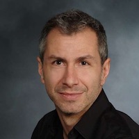
Simon Scheuring is Professor of Physiology and Biophysics in Anesthesiology and the head of the “Bio-AFM-Lab” at Weill Cornell Medicine, associated to the ‘Department of Anesthesiology’ and the ‘Department of Physiology & Biophysics’.
Simon Scheuring is a trained biologist from the Biozentrum at the University of Basel, Switzerland (1992-1996). During his PhD in the laboratory of Andreas Engel (1997-2001), he learned electron microscopy and atomic force microscopy, and got interested in membrane proteins. During this period he worked on the structure determination of aquaporins in collaboration with Peter Agre. During his postdoc at the Institut Curie in Paris, France, in the laboratory of Jean-Louis Rigaud (2001-2004), he learned membrane physical chemistry and developed atomic force microscopy for the study of native membranes. As a permanent researcher (2004-2007) and junior research director (2007-2012) he started to set up his lab at the Institut Curie in Paris, France. He then moved to Marseille (2012-2016) where he built a larger independent laboratory named “Bio-AFM-Lab” administrated by INSERM and Université Aix-Marseille. He got promoted senior research director of INSERM (2012). For January 2017, Simon Scheuring moved to Weill Cornell Medicine (WCM) in Manhattan, New York, USA (2017), where he accepted a Professor position and opened a new laboratory focusing on the development and application of atomic force microscopy based technologies for the study of membrane phenomena such as the structure, assembly, dynamics and conformational changes of channels, transporters, receptors and membrane remodeling proteins. His objective is to head a dynamic young research team with members from different scientific fields ranging from biology, physics and engineering to develop and use the atomic force microscopy to provide novel insights into the processes taking place in biological membranes.

