Have a very Nano New Year with the Molecular Foundry’s 2024 Calendar, featuring colorful submissions from the 2023 NanoArt Image Contest. The work behind these images comes from Foundry staff and users spanning all seven of the Foundry’s technical facilities.
Do you know which day is ‘DNA Day’? Check the Foundry calendar!
Download and print your own copy.
About the 2024 Calendar
January
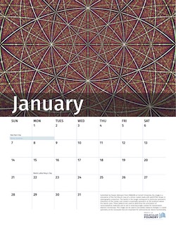
Submitted by Steven Zeltmann from PARADIM at Cornell University, the image is a simulation of the full Kikuchi map of a silicon crystal made with py4DSTEM, shown in stereographic projection. The bands in the image correspond to particular symmetric directions of the crystal, and crystal is maximally symmetric at the locations where the bands cross. These patterns are used to determine the orientation of nanocrystalline materials and to aid in orienting single crystals for transmission electron microscopy. This image can be used to accurately measure changes in crystal symmetry at the nanoscale that are important for semiconductor device functionality.

February
Submitted by Stephanie Ribet from the Foundry’s NCEM facility, this STEM image, taken by the TEAM 0.5 microscope, shows core shell nanoparticles arranged in a heart. High resolution imaging with electron microscopy techniques can be used to establish structure-property relationships in nano materials. They investigated the structure of these core shell nanoparticles synthesized at the Foundry with STEM techniques.

March
Submitted by Aidar Kemelbay and created in work by Aidar Kemelbay, Fabrizio Riminucci, Marco D’Alessandro from the Molecular Foundry in the Foundry’s Nanofabrication facility. In celebration of National Nano Day, they prepared a Bragg cake with a polymer cherry on top. This structure was baked in oven at 650C in hydrogen disulfide to obtain transition metal dichalcogenide based all-dielectric Bragg reflector.

April
Submitted by Tev Kuykendall from work by Tev Kuykendall and Alexander Herman in the Inorganic Facility, this image shows lithographically defined lines of 2D transition metal dichalcogenides (TMDs), made using a Lateral Conversion synthesis method. TMDs have many applications, including optical, electronic and chemical sensing devices.
May
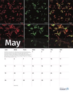
Submitted by Bruce Cohen from the Biological Nanostructures facility, this image shows amyloid beta peptide plaques like those found in Alzheimer’s brain, stained with new Foundry fluorophore Phazr (left, red), standard probe ThT (center, green), and merged images (right). Phazr fluorescence of plaques is both brighter and more extensive than with standard ThT probes, even at much lower concentrations. This was collected as part of work by Zeming Wang, Line G. Kristensen, Luis A. Valencia, Kyleigh L. Range, Yen H. Ho, Behzad Rad, Corie Y. Ralston & Bruce E. Cohen.
June
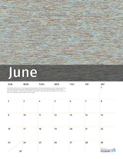
Submitted by Emory Chan from the Inorganic Facility. This image shows the evolution of the excited states of erbium dopants over time in an upconverting nanoparticle simulated with kinetic Monte Carlo. Each row represents one of the 2220 erbium ions doped in a sodium yttrium fluoride nanocrystal. The colors depict the energy of each erbium ion over time, moving across the rows from left to right.
July
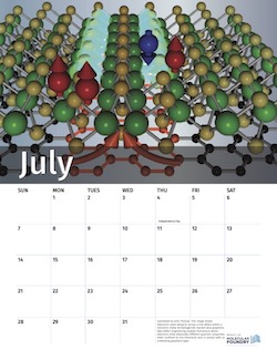
Submitted by John Thomas from work in the Imaging Facility. This image shows electronic-state behavior across a line defect within a transition metal dichalcogenide stacked atop graphene. New defect engineering enables formations where electrons show drastically different quantum properties when confined to one dimension and in contact with an underlying graphene layer.
August
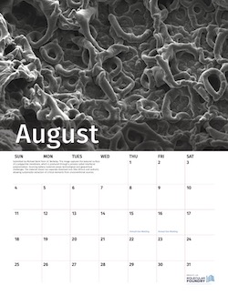
Submitted by Michael Baird from UC Berkeley from work that utilized the Organic and Imaging facilities. This image captures the textured surface of a polypyrrole membrane, which is produced through a process called interfacial polymerization. Sourcing battery materials poses technological and geopolitical challenges. The material shown can separate dissolved ions (like lithium and sodium), allowing sustainable extraction of critical elements from unconventional sources.
September
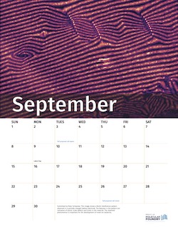
Submitted by Peter Schweizer and collected from work conducted at NCEM. This image shows a Moiré interference pattern observed in a partially charged battery electrode. The features in the pattern are indicative of atomic scale defects and strain in the material. The observed phenomenon is important for the development of novel ion batteries.
October
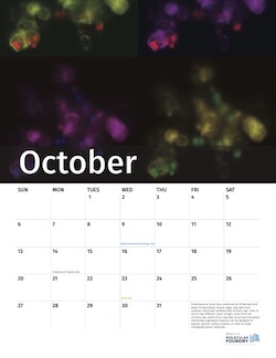
Diatomaceous Soup Cans, produced by Ed Barnard and Adam Schwartzberg in the Imaging and Nanofabrication facilities. Diatom algae cells with their surfaces chemically modified with mCherry dye. Color is due to two different colors of light, some from the mCherry dye, others from naturally occurring chlorophyll. Genetically engineered diatoms can be designed to express specific surface proteins in order to make biologically grown materials.

November
Submitted by Beihang Yu from work in the Nanofabrication facility. A “boat” in a DNA origami sea. Aluminum oxide is first selectively grown inside the DNA origami to replicate its structure, then all organics are removed leaving behind the aluminum oxide as a mask. We use the mask to transfer the “DNA shape” into the Si substrate. The “boat” is an impurity particle spotted under SEM. This is a workflow that we can use to leverage biomacromolecules, which are highly programmable and precise, as templates for high-precision pattern transfer.

December
Submitted by Stephanie Ribet, this image was selected as the winner of the 2023 NanoArt Image Contest. This image, taken at NCEM, shows a converged beam electron diffraction pattern of a twisted bilayer of TaSe2. Twisted bilayers of 2D materials are of great interest due to unusual physical, electronic, and optical properties that arise from the complex bilayer stacking. This TaSe2 stack forms a Moiré pattern, which we can study with electron ptychography and interferometry.

