The Molecular Foundry hosted it’s annual NanoArt competition in celebration of National Nano Day – 10/9 – which helps raise awareness of nanotechnology, how it is currently used in products that enrich our daily lives, and the challenges and opportunities it holds for the future.
We had a number of great entries from the Molecular Foundry community, and now the votes are in! The top entries will be professionally printed and displayed in the Molecular Foundry’s lobby.
1st Place

“Sunset in the Atomic Alps”, submitted by Hudson Shih as part of work by Hudson Shih, Seung Sae Hong, and Yayoi Takamura from UC Davis – Atomic terrace steps on the (001) facet of a single-crystal membrane of strontium cobalt oxide (Sr2Co2O5 ). These steps arise due to a small angle of miscut relative to the facet (<0.5 °). If the miscut angle were exactly 0 degrees, no steps would be present! These single crystal oxide membranes provide an ideal material platform for plan-view transmission electron microscope (TEM) imaging at the National Center for Electron Microscopy because they have millimeter lateral dimensions, but nanometer scale thicknesses. With TEM of Sr2Co2O5 membranes, oxygen ion diffusion can be observed within the crystal lattice.
2nd Place
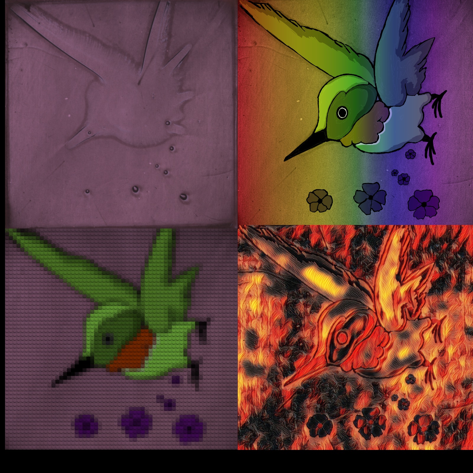
Submitted by Thong M Nguyen from the University of Washington. Hummingbird species deposited using AutoBot. The hummingbird shape is a consequence of a non-ideal solution engineering step during halide perovskite thin film deposition on the AutoBot robotic platform. Halide perovskite materials are promising for optoelectronics including photovoltaics, LEDs, spintronic applications.
Part of work involving Thong M Nguyen (UW), Ansuman Halder, Shijing Sun (UW), and Carolin Sutter-Fella
3rd Place
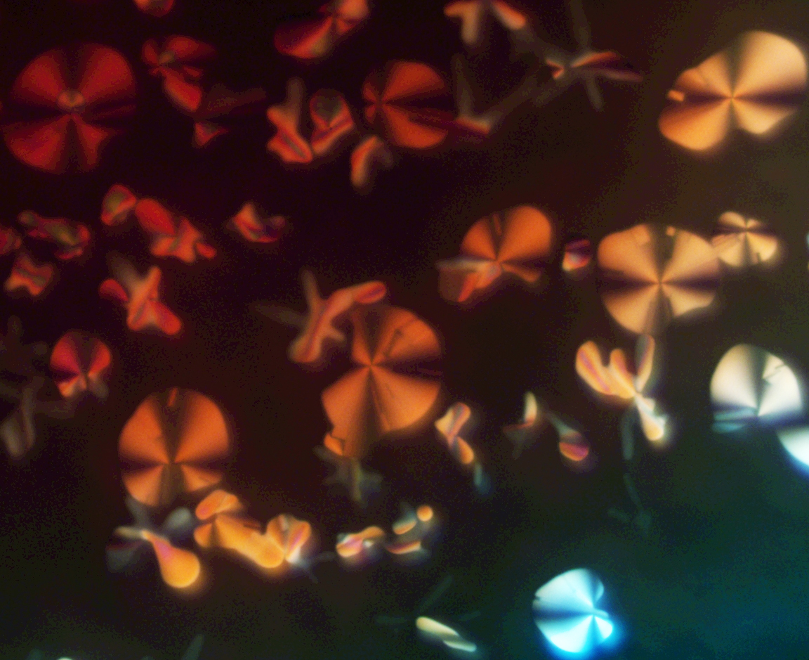
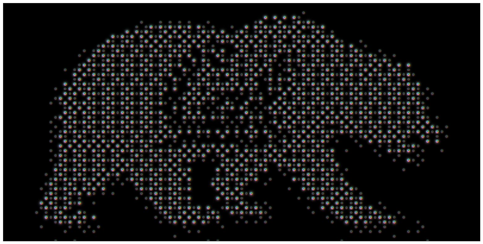
Submitted by Yi Liu. Nanobutterflies: birefringence patterns of liquid crystalline materials captured under an optical microscope, resembling the vivid color and shape of butterflies. Organic liquid crystals are useful for flexible displays and nano-optics.
4th Place
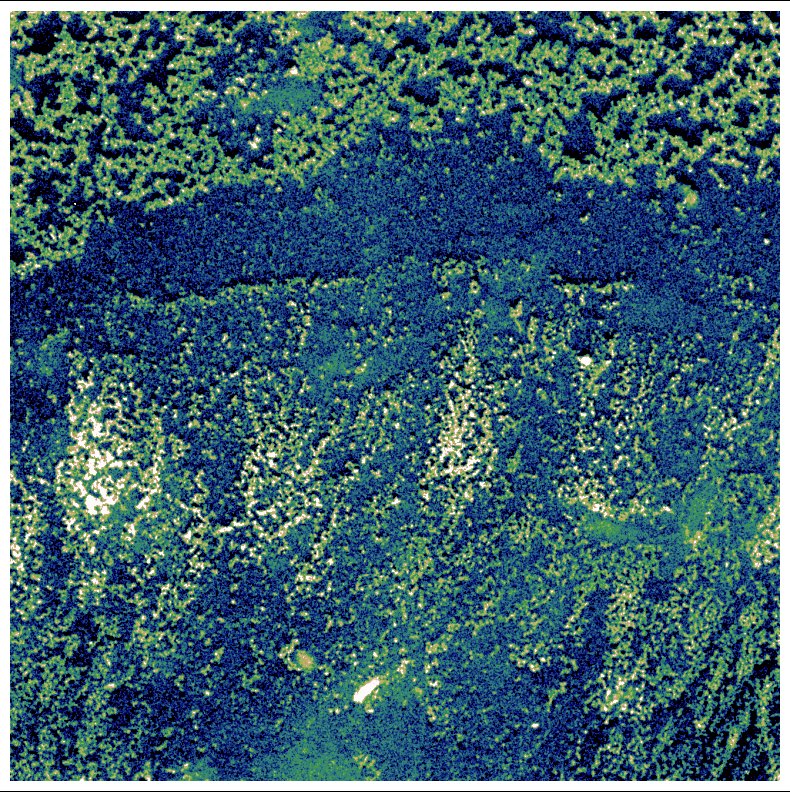
Submitted by Min Gee Cho. This TEM image captures the assembly of 10 nm cobalt oxide nanocrystals. The expansive field of view unveils an intricate landscape reminiscent of beautiful undersea terrain. The contrasting assemblies of nanoparticles in the upper and lower regions are shaped by the liquid flow of micro-droplets during evaporation, guiding their arrangements on the carbon substrate. This natural phenomenon, born from the delicate balance of micro-liquid flow, embodies the harmonious interaction of local forces at the nanoscale. The visual resemblance to terrestrial topography explores the hidden beauty of the nanoscale world—naturally occurring patterns that express nature’s artistry in a realm invisible to the naked eye. (Microscope: Tecnai / Field-of-view: 7900 nm) The overall project is examining the self-assembly of nanoparticles for complex higher-order nanostructures.
5th Place
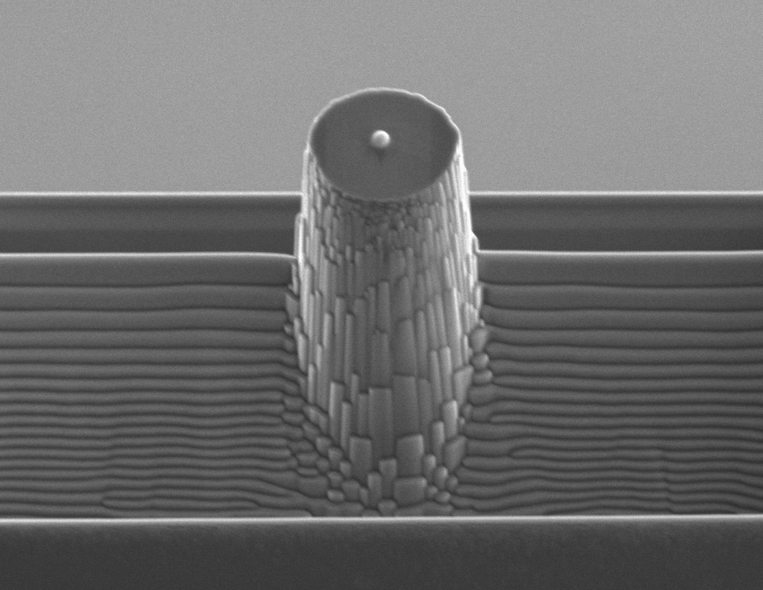
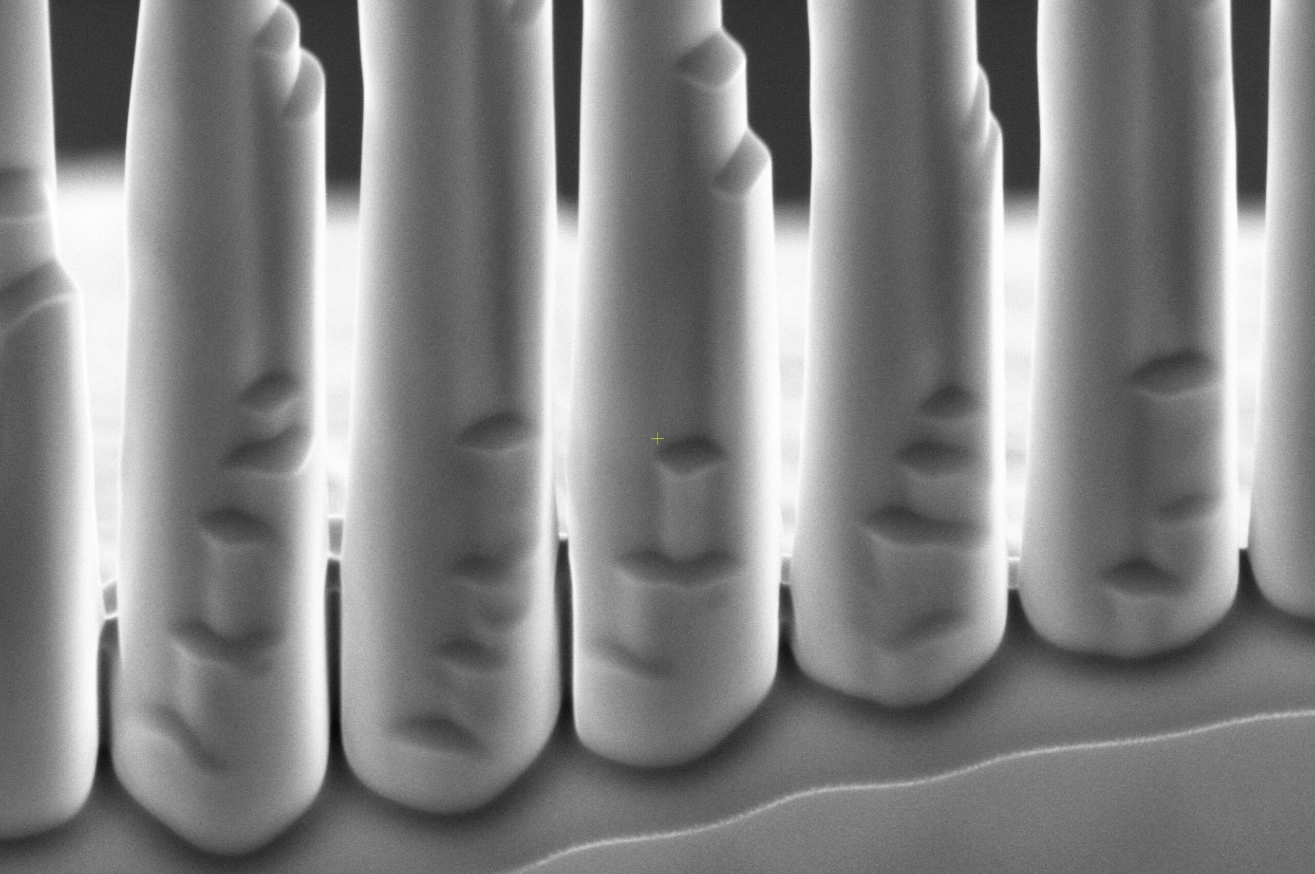
Submitted by Iliana Marrujo. A diamond pillar with 500nm platinum bead for mechanical testing. Diamond Anvil Cells (DACs) are used to investigate the behavior of materials under extreme pressure and observe pressure-induced phase transformations. The researchers are developing methods to visualize these processes using transmission electron microscopy.
6th Place

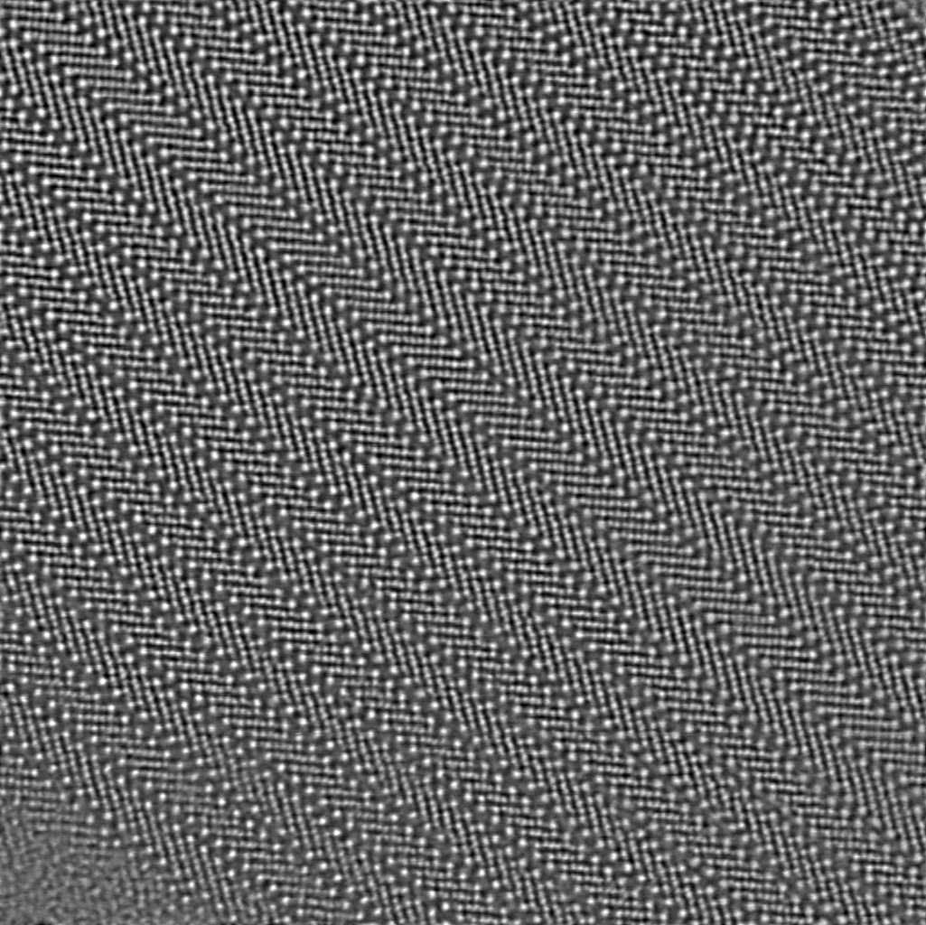
Submitted by Stephanie Ribet. Four-dimensional scanning transmission electron microscopy (4D-STEM) can be used to map the phase and orientation of materials on the nanoscale. This image shows a semi-crystalline polymer blend, where the colors represent phase and the direction of the lines shows the orientation of crystalline grains. This sustainability study was targeted at understanding the structure of polymers with different functionalization. Characterizing the nanoscale morphology allows us to to design better materials and methods for plastic upcycling.
The image was taken as part of work by Stephanie Ribet (MF), Colin Ophus (Stanford), Karen Bustillo (MF), Eliza Neidhart (UNC), Frank Leibfarth (UNC), Mutian Hua (MF), and Brett Helms (MF).
Honorable Mentions

