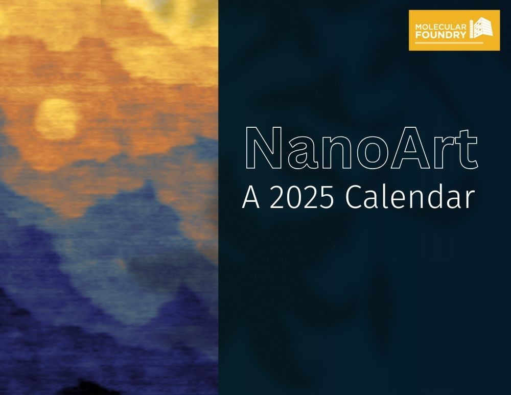
Have a very Nano New Year with the Molecular Foundry’s 2025 Calendar, featuring colorful submissions from the 2024 NanoArt Image Contest. The work behind these images comes from Foundry staff and users spanning all seven of the Foundry’s technical facilities.
Do you know which day is ‘National Nano Day’? Check the Foundry calendar!
Download and print your own copy.
About the 2025 Calendar

January
A ‘nano-onion’: Using liquid-phase transmission electron microscopy, we discovered a new covalent organic framework (COF) ‘onion’ nanostructure with significant structural flexibility, including variations in orientation and curvature. By mapping the onion layers in different colors, we revealed the disorder and defects within the framework. The COF onion shows great potential for carbon capture, and notably, this is the first observation of its onion structure at the molecular level. The image was taken using the THEMIS microscope at NCEM by Qi Zheng and Haimei Zheng from the Materials Sciences Division at Lawrence Berkeley National Lab.
February
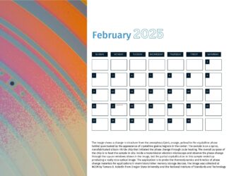
The image shows a change in structure from the amorphous (pink, orange, yellow) to the crystalline phase (white) punctuated by the appearance of crystalline grains/regions in the center. The sample is on a spiral, nanofabricated silicon nitride chip that initiated the phase change through Joule heating. The overall purpose of the chip is to heat the sample in-situ inside a transmission electron microscope and observe the phase change through the square windows shown in the image, but the partial crystallization in this sample ended up producing a really nice optical image. The application is to probe the thermodynamics and kinetics of phase change materials for applications in memristors/other memory storage devices. The image was collected at NCEM by Tamara D. Koledin from Oregon State University and the National Institute of Standards and Technology
March
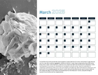
For the first time we found that the as-yet-enigmatic compounds found in some coral larvae in huge amounts can form rose-like crystalline aggregates. Similarly to thorns in roses, these compounds protect their owner being toxic to other corals. Montiporic acids are rare compounds with triple bonds found in Montipora corals. Learning about the process of crystallization of montiporic acids will uncover how the corals thrive in competitive environment of the corals reefs, the hotspots of biodiversity. Apart from that this compound is not toxic just for the competing species but also for cancer cells and thus they may be used in human medicine. This image was taken by Jana Pilatova and Behzad Rad from the Molecular Foundry using the Biological Nanostructures facility and the Imaging facility.
April
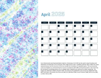
Four-dimensional scanning transmission electron microscopy (4D-STEM) can be used to map the phase and orientation of materials on the nanoscale. This image shows a semi-crystalline polymer blend, where the colors represent phase and the direction of the lines shows the orientation of crystalline grains. This sustainability study was targeted at understanding the structure of polymers with different functionalization. Characterizing the nanoscale morphology allows us to to design better materials and methods for plastic upcycling. The image was collected at NCEM using the TEAM I microscope and py4DSTEM as part of work by Stephanie Ribet (MF), Colin Ophus (Stanford), Karen Bustillo (MF), Eliza Neidhart (UNC), Frank Leibfarth (UNC), Mutian Hua (MF), and Brett Helms (MF).
May
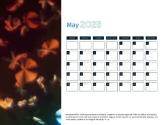
Nanobutterflies: birefringence patterns of liquid crystalline materials captured under an optical microscope, resembling the vivid color and shape of butterflies. Organic liquid crystals are useful for flexible displays and nano-optics. Created in the Organic facility by Yi Liu.
June
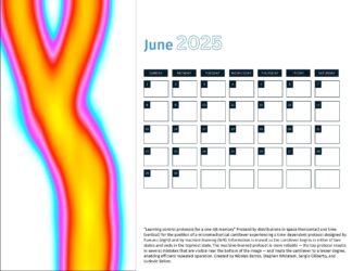
“Learning control protocols for a one-bit memory”
Probability distribution in space (horizontal) and time (vertical) for the position of a micromechanical cantilever experiencing a time-dependent protocol developed by machine learning. Information is erased as the cantilever begins in either of two states (top) and ends in the rightmost state (bottom). The machine-learned protocol is more reliable than our human-designed protocols, and heats the cantilever to a lesser degree, enabling efficient repeated operation. The protocol was trained in the Theory facility and deployed in experiments at ENS Lyon by Nicolas Barros, Stephen Whitelam, Sergio Ciliberto, and Ludovic Bellon.
July
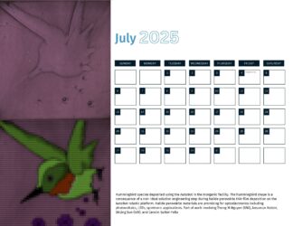
Hummingbird species deposited using the AutoBot in the Inorganic facility. The hummingbird shape is a consequence of a non-ideal solution engineering step during halide perovskite thin film deposition on the AutoBot robotic platform. Halide perovskite materials are promising for optoelectronics including photovoltaics, LEDs, spintronic applications. Part of work involving Thong M Nguyen (UW), Ansuman Halder, Shijing Sun (UW), and Carolin Sutter-Fella.
August
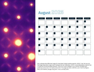
This a selected area diffraction image of a disordered rocksalt cathode material, LMTOF, in the 100 zone axis. The image shows short range order in between the main reflections. LMTOF is a promising battery material that is comprised of earth abundant elements. This can be used to mitigate scarce element shortages from producing commercial Li ion battery chemistries. The image was taken at NCEM as part of work by Meghan Shen, Karen Bustillo, Chengyu Song, John Turner, and Mary Scott.
September
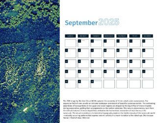
This TEM image by Min Gee Cho at NCEM captures the assembly of 10 nm cobalt oxide nanocrystals. The expansive field of view unveils an intricate landscape reminiscent of beautiful undersea terrain. The contrasting assemblies of nanoparticles in the upper and lower regions are shaped by the liquid flow of micro-droplets during evaporation, guiding their arrangements on the carbon substrate. This natural phenomenon, born from the delicate balance of micro-liquid flow, embodies the harmonious interaction of local forces at the nanoscale. The visual resemblance to terrestrial topography explores the hidden beauty of the nanoscale world—naturally occurring patterns that express nature’s artistry in a realm invisible to the naked eye. (Microscope: Tecnai / Field-of-view: 7900 nm)

October
“Sunset in the Atomic Alps”, by Hudson Shih, Seung Sae Hong, and Yayoi Takamura from UC Davis. This image won first place in the 2024 NanoArt Image contest.
Atomic terrace steps on the (001) facet of a single-crystal membrane of strontium cobalt oxide (Sr2Co2O5). These steps arise due to a small angle of miscut relative to the facet (<0.5 °). If the miscut angle were exactly 0 degrees, no steps would be present! These single crystal oxide membranes provide an ideal material platform for plan-view transmission electron microscope (TEM) imaging at the National Center for Electron Microscopy because they have millimeter lateral dimensions, but nanometer scale thicknesses. With TEM of Sr2Co2O5 membranes, oxygen ion diffusion can be observed within the crystal lattice.
November
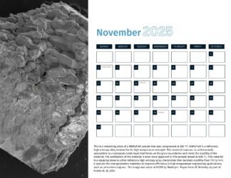
This is a remaining piece of a NbMoTaW sample that was compressed at 800 °C. NbMoTaW is a refractory high-entropy alloy desired for its high temperature strength. This material however, is unfortunately susceptible to a nanoscale oxide layer that forms on the grain boundaries and limits the ductility of the material. The oxidization of the material is even more apparent in this sample tested at 800 °C. This material is a stepping stone to other refractory high-entropy alloy chemistries that maintain ductility from 77K to 1473 K and are the next generation materials to improve efficiency in high temperature engineering applications, such as jet turbine engines. The image was taken at NCEM by Madelyn I. Payne from UC Berkeley as part of Kumar et. al, 2024.
December
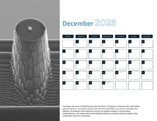
This image was taken at NCEM by Iliana Marrujo from UC Berkeley. A diamond pillar with 500nm platinum bead for mechanical testing. Diamond Anvil Cells (DACs) are used to investigate the behavior of materials under extreme pressure and observe pressure-induced phase transformations. The researchers are developing methods to visualize these processes using transmission electron microscopy.

